Microchip CE-MS
Since CE is an ultra-low volume separation technique it has been an obvious development to execute CE in narrow conduits on microfluidic chips (microchip capillary electrophoresis, MCE). Since early 2000 MCE has been deployed as an instrumental separation method in dedicated devices such as the Bio-analyzer (Agilent Technologies) or the LabChip (Caliper Technologies, now Perkin Elmer). In contrast though to general purpose CE instrumentation, these devices have highly dedicated tasks like DNA and protein sizing and quantitation. Coupling of such devices with MS is prohibited by their design. Therefore, coupling MCE with MS has remained an area of research since the mid-nineties [1-2].
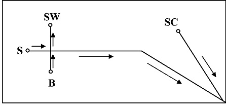
Despite the great potential, no commercial solution for MCE-MS is available on the market. As one example of this work, the approach by Michael Ramsey et al. is given here [44-46]. The principle layout of their chip is shown in figure 7.

MCE-MS according to Ramsey et al
The chip is prepared from 150-µm thick Corning 0211 borosilicate glass. The channels that were obtained were about 75 µm wide and 10 µm high. The length of the separation channel S was 4.7 cm. The side channel was 2 cm in length. The side channel is intended to deliver an EOF-driven sheath solvent and connects to the separation channel 200 µm before the tip. In Ramsey's initial work, the separation channel was coated with a cationic polymer. This rendered the surface of the channel positively charged and gave a strong anodic EOF and suppressed adsorption of peptides and proteins with low pH BGE. The side channel was not coated and delivered a small cathodic EOF. In this way, the flow direction in both channels is the same. To run the chip 1 kV was applied to SW and 4kV to SC (to generate a cathodic EOF and the ES voltage). Given that the mass spectrometer inlet was at the ground (Micromass Q-ToF) the circuit is closed. BGE was 0.2% acetic acid in methanol/water (1:1). Peptides, proteins, and a BSA digest were used as samples. A moderate sensitivity was obtained, i.e. around 1 µM in sample concentration.
![]()
[1] Breadmore M.C. (2012) J. Chromatogr. A 1221 42-55
[2] Felhofer J.L., Blanes L, Garcia C.D. (2010) Electrophoresis 31 2469-2486
[3] Mellors J.S., Gorbounov V, Ramsey R.S., Ramsey J.M. (2008) Anal Chem 80 6881-6887
[4] Batz N.G., Mellors J.S., Alarie J.P., Ramsey J.M. (2014) Anal Chem 86 3493-3500
[5] Mellors J.S., Black W.A., Chambers A.G., Starkey J.A., Lacher N.A., Ramsey J.M. (2013) Anal Chem 85 4100-4106
[6] Mellors J.S., Jorabchi K, Smith L.M., Ramsey J.M. (2010) Anal Chem 82 967-973
